


ttp://www.zo.utexas.edu/faculty/sjasper/bio301L/digestion.html
Alimentary canal in man


In humans and other vertebrates , the digestive system consists of a tubular gastro intestinal tract and accessory digestive organs. This is a one way transport of food.
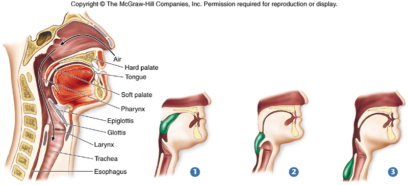
Mouth opens to the buccal cavity .The pharynx which is the common passage of the oral and nasal cavities .The pharynx leads to the oesophagus, a muscular tube ,delivers food to the stomach.
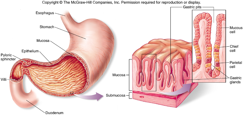
From the stomach food passes to the first part of intestine where the digestive enzymes continues the digestive process.
The products then pass across the wall of the small intestine into the blood stream.
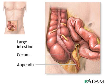
Then the remains into the large intestine where water and minerals are absorbed.
The waste products then enter to the rectum and expelled through the anus.
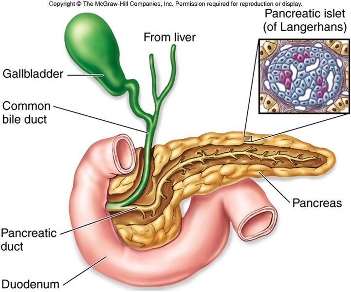
The accessory organs are the liver - produce bile gall bladder – stores and concentrates bile pancreas - produces pancreatic juice which contain digestive enzymes and bicarbonates
Both bile and pancreatic juice first secreted into the first region of the small intestine and aid digestion
Human like all placental mammals lack a cloaca and have a seperate exit from the digestive tract through rectum and anus

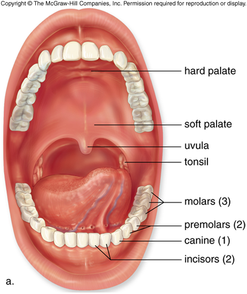
Mouth
Humans are omnivorous, are specialized for eating both plants and animal food
Humans are omnivorous in front and herbivorous in back of the mouth.
There are four types of teeth in the mouth.
Incisors- Four front teeth in the upper and lower jaws .
They are sharp ,chaise – shaped for biting
Canine - Each side of the incisors are sharp pointed teeth called
canine for tearing food .
Pre molars- behind pre molars are canine for crushing food.
Molar – with flattened rigid surfaces for grinding and crushing food
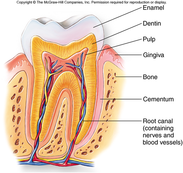
Each tooth is alive with a central pulp containing nerves and blood vessels.
The actual chewing surface is a hard layered enamel layer over the softer dentin which forms the body of the tooth.

Saliva
Inside the mouth tongue mixes food with a mucous solution , saliva. pH is 6.5 – 7.5
In humans three pairs of salivary glands, secreted saliva into mouth through ducts.
Saliva is a mixture of secretions of salivary glands and mucous glands of the buccal cavity epithelium.
The secretion of the salivary glands are controlled by the nervous system.
The presence of the food in the mouth triggers an increased rate of secretion.
Functions
Saliva moisten and lubricates the food and this is easier to swallow the food .
Water contained in saliva wet the dry food.
Ions in saliva such as K+ , Cl -
It contains hydrolytic enzyme, salivary amylase. Cl - activates salivary amylase and converts starch into maltose.
Polysaccharides salivary amylase Disaccharides
Starch Maltose
Mucus in saliva lubricates food.
Lysozymes present in saliva destroy bacteria.
Saliva helps in taste reception.
Secretion of saliva is stimulated by parasympathetic nervous system and inhibited by sympathetic nervous system.
Ingested food is fragmented through the tearing or grinding action of specialized teeth. Series of pharyngeal muscle contractions begins and then food mixed with saliva is swallowed and enters the esophagus. Saliva does not abrade the tissue , it passes on its way through the esophagus.
Esophagus
Swallowed food enters the esophagus , which connects the
pharynx to the mouth .
The upper third is enveloped to skeletal muscle, for
voluntary control of swallowing.
The lower two third is surrounded by involuntary smooth
muscle.
The food moves along
the esophagus to the stomach by
peristalsis movements.
The food moves down to the stomach is controlled by a ring of
circular smooth muscle or a sphincter ,
that opens in response to the pressure extend by the food.
Contraction of this sphincter prevents food in the stomach
from moving back into the esophagus. The
human esophagus is closed off except
during swallowing.

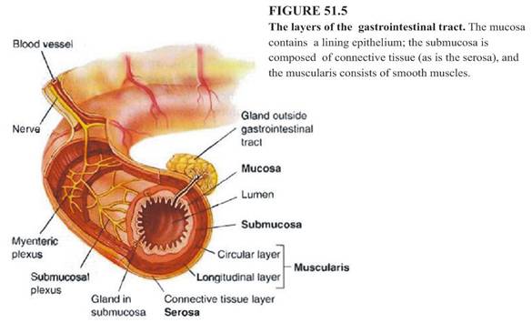
Basic plan consists of four main layers of the gut wall.
From outermost layer to innermost layer are,
Serosa – Consists of fibrous connective tissue and are covered the external surface
of the tract with peritonium.
Muscularis – Just outside the sub mucosa. Consists of double layer of smooth
muscles.The muscles in the outer layer arranged longitudinally and
the muscles in the inner layer have a circular muscles.
Auerbach’s plexus / nerve plexus are present between the
longitudinal muscle layer and the circular muscle layer.
Sub Mucosa –Consists of loose connective tissue .
Meissner’s plexus are present between sub mucosa and circular muscle
layer.
Nervous intertwined regions are called plexus, are located in the sub mucosa, help to regulate gastro intestinal activities
Mucosa - Consists of loose connective tissue , lined with an epithelium.
Major variations are those seen in the basic plan occur in the mucosa.
They are associated with particular functions.
Tissue plan in small intestine
1.Outermost layer is serosa
2.The layer near to the outermost layer
is muscularis.
Circular muscle layer is thicken.
Circular muscle layer is thicken.
3.Next inner layer is submucosa.Bruner’s glands are
present in the sub mucosa.These glands secret the
mucous alkaline secretion that nutralize the chyme and
protects the deuodenum wall from the damage.
present in the sub mucosa.These glands secret the
mucous alkaline secretion that nutralize the chyme and
protects the deuodenum wall from the damage.
4.Innermost layer is mucosa which
consists number of
circular foldings called Liberkun cripts . This increase the
surface area of the small intestine.
circular foldings called Liberkun cripts . This increase the
surface area of the small intestine.
5.The small intestine consists mucosa,
water, enterokinase
and enteropeptidase.
and enteropeptidase.
6.Presence of villi.
7.Intestinal glands among the villi
that are called enterocytes secretes entero gasteron.
8.Lymph noduleswhich are called Payers
patches that helps to produce antibodies
9.Presence of network of blood capillaries.
10. Network of lymph vessels – Lacteals
11.Cells in the inner lining / epithelium have micro villi

http://science.waltermack.com/smack/yr8biol/digestion2.gif


http://glencoe.mcgraw-hill.com/sites/9834092339/student_view0/chapter48/